Blood Vessels Labeled Brain : Arterial Supply Of The Brain Circle Of Willis Geeky Medics : Identify all of the blood vessels that are illustrated in the figure as you can while holding or otherwise examining whole brain specimens.
Blood Vessels Labeled Brain : Arterial Supply Of The Brain Circle Of Willis Geeky Medics : Identify all of the blood vessels that are illustrated in the figure as you can while holding or otherwise examining whole brain specimens.. In this video i discuss the major arteries that supply the brain, starting with the internal carotid and vertebral arteries and covering many of the major. They do not have muscle layers and allow the exchange of substances between the blood and the a few structures (such as cartilage and the lens of the eye) do not contain blood vessels and are labeled avascular. Blood vessels in red in close communication with proliferating neuronal cells in the mouse cortex at embryonic day 10. Blood travels from the heart in arteries, which branch into smaller and smaller vessels, eventually becoming arterioles. This vessel supplies blood to the front part of your brain, knows as your frontal lobe.
Blood is supplied to the brain through 2 major pairs of arteries. The brain and its surrounding blood vessels exist in a close relationship. Diseases of the brain and nervous system(a health education guide): The two cell types ensure the integrity of the neural vasculature by maintaining the blood. Label the veins of the anterior forearm and hand.
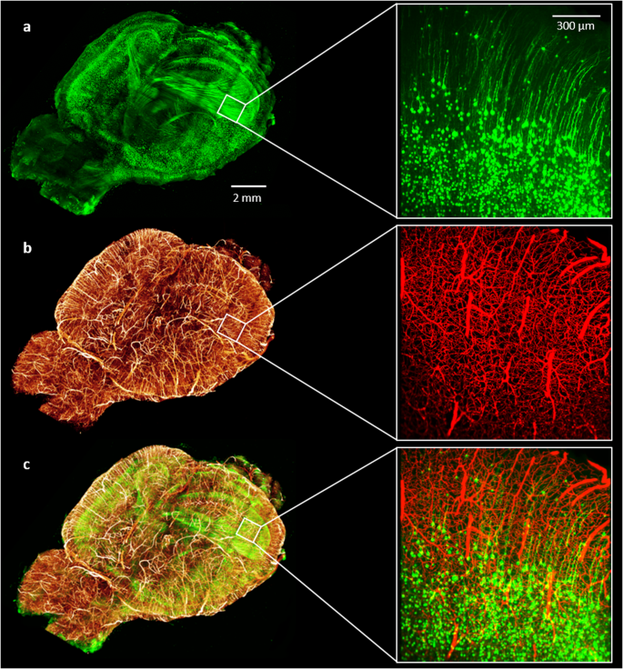
Whole Brain Vasculature Reconstruction At The Single Capillary Level Scientific Reports from media.springernature.com In this video i discuss the major arteries that supply the brain, starting with the internal carotid and vertebral arteries and covering many of the major. The two cell types ensure the integrity of the neural vasculature by maintaining the blood. Blood vessels are referred to collectively as the vascular system and, together with the heart, make up the circulatory system or cardiovascular system. Only some of the vessels that exist in a real brain have been labeled. What we see here are the blood vessels that extend along the inferior surface of the temporal and occipital lobe. Cerebral small vessel disease • not a single disease • group of diseases with different pathologies and different aetiologies • affecting the small arteries, arterioles, venules. | study material, lecturing notes, assignment mr angiography makes the use of the mri magnetfor the examination of the blood vessels in this test no catheter is required to be introduced in the blood. Blood vessels are intricate networks of hollow tubes that transport blood throughout the entire body so that it can deliver valuable nutrients to and remove waste from cells.
Supplies the anterior brain and the vertebral a.
Blood travels from the heart in arteries, which branch into smaller and smaller vessels, eventually becoming arterioles. The vessels of the brain circulate blood throughout the brain to ensure that all of its nerves and cells receive the nutrients they need. Label the blood vessels of the male pelvis using the hints provided. Towards the anterior side of the brain, those arteries are the internal carotid arteries. Cerebral small vessel disease • not a single disease • group of diseases with different pathologies and different aetiologies • affecting the small arteries, arterioles, venules. (a) at 3 days after stroke. The brain and its surrounding blood vessels exist in a close relationship. Very thin blood vessels found in the middle of tissue and organs. Endothelial cells are labeled in red and pericytes in green. The brain can autoregulate blood flow in order to ensure constant flow that is isolated from the arterial blood supply to the brain can be divided into the anterior and posterior circulation. Researchers have discovered how cells of the blood vessels sense the metabolic condition of the brain and alter vascular function in response. This vessel supplies blood to the front part of your brain, knows as your frontal lobe. In the article on the ventricles within the cns, we will discuss their structure and.
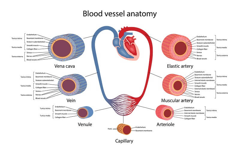
They do not have muscle layers and allow the exchange of substances between the blood and the a few structures (such as cartilage and the lens of the eye) do not contain blood vessels and are labeled avascular. Blood vessels are tubes that run through the transport system in which blood is transported. Blood travels from the heart in arteries, which branch into smaller and smaller vessels, eventually becoming arterioles. These vessels transport blood cells, nutrients, and oxygen to the tissues of the body. Blood vessels are referred to collectively as the vascular system and, together with the heart, make up the circulatory system or cardiovascular system.
Circulatory System The Definitive Guide Biology Dictionary from biologydictionary.net Blood travels from the heart in arteries, which branch into smaller and smaller vessels, eventually becoming arterioles. Towards the anterior side of the brain, those arteries are the internal carotid arteries. However, they have observed blood vessel damage caused. | study material, lecturing notes, assignment mr angiography makes the use of the mri magnetfor the examination of the blood vessels in this test no catheter is required to be introduced in the blood. Supplies the posterior brain, blood supply to the entire brain is ensured by anastomoses between the vessels. The 500 ms patients, both adults and children, also underwent mri scans of the brain to measure iron deposits in surrounding areas of the brain. Cerebral small vessel disease • not a single disease • group of diseases with different pathologies and different aetiologies • affecting the small arteries, arterioles, venules. The brain and its surrounding blood vessels exist in a close relationship.
They do not have muscle layers and allow the exchange of substances between the blood and the a few structures (such as cartilage and the lens of the eye) do not contain blood vessels and are labeled avascular.
Towards the anterior side of the brain, those arteries are the internal carotid arteries. • identification of blood vessels as arteries, capillaries or veins from the structure of their walls. In this video i discuss the major arteries that supply the brain, starting with the internal carotid and vertebral arteries and covering many of the major. The two cell types ensure the integrity of the neural vasculature by maintaining the blood. Blood is supplied to the brain through 2 major pairs of arteries. | study material, lecturing notes, assignment mr angiography makes the use of the mri magnetfor the examination of the blood vessels in this test no catheter is required to be introduced in the blood. The carotid arteries and the vertebral arteries anterior cerebral artery (aca): The brain and its surrounding blood vessels exist in a close relationship. Traditionally, pais has been explained as being caused by a blood clot forming within the ageing placenta, entering the fetal circulation, embolising across the patent foramen ovale, travelling into the left ventricle, into the ascending aorta and then one of the main three branches of the thoracic aorta. Blood supply to the brain is supplied by two main pairs of arteries, the internal carotid arteries and the vertebral arteries. Cerebral small vessel disease • not a single disease • group of diseases with different pathologies and different aetiologies • affecting the small arteries, arterioles, venules. However, they have observed blood vessel damage caused. The dense tight junctions between endothelial cells prevent paracellular transport through the.
Blood is supplied to the brain through 2 major pairs of arteries. Researchers have discovered how cells of the blood vessels sense the metabolic condition of the brain and alter vascular function in response. • identification of blood vessels as arteries, capillaries or veins from the structure of their walls. Blood vessels and lymph nodes. | study material, lecturing notes, assignment mr angiography makes the use of the mri magnetfor the examination of the blood vessels in this test no catheter is required to be introduced in the blood.
Heart Anatomy Anatomy And Physiology from s3-us-west-2.amazonaws.com At the same time, blood vessel double labeled for brdu and reca were present in remote areas relative to the stroke lesion (figure 3h , arrows) and in the figure 4. In this video i discuss the major arteries that supply the brain, starting with the internal carotid and vertebral arteries and covering many of the major. This is particularly important structure due to its clinical implications, which are discussed in more detail in the article. He says the restricted vessels prevent the blood from draining fast enough and injure the brain by causing a build up of iron which leads to ms. Comes off the subclavian a., ascends although the internal carotid a. Blood supply to the brain is supplied by two main pairs of arteries, the internal carotid arteries and the vertebral arteries. The difference in the structural characteristics of arteries, capillaries and veins is attributable to their respective functions. (a) at 3 days after stroke.
Comes off the subclavian a., ascends although the internal carotid a.
Very thin blood vessels found in the middle of tissue and organs. However, they have observed blood vessel damage caused. They also take waste and carbon dioxide away from the tissues. Function and homeostasis of the brain relies on communication between its complex network of cells. The blood vessels are the components of the circulatory system that transport blood throughout the human body. The 500 ms patients, both adults and children, also underwent mri scans of the brain to measure iron deposits in surrounding areas of the brain. What we see here are the blood vessels that extend along the inferior surface of the temporal and occipital lobe. They do not have muscle layers and allow the exchange of substances between the blood and the a few structures (such as cartilage and the lens of the eye) do not contain blood vessels and are labeled avascular. Cerebral small vessel disease • not a single disease • group of diseases with different pathologies and different aetiologies • affecting the small arteries, arterioles, venules. Blood is also supplied to the brain by the vertebral a. Researchers have discovered how cells of the blood vessels sense the metabolic condition of the brain and alter vascular function in response. Microscopically, it is formed by the endothelium of the blood vessel. The brain can autoregulate blood flow in order to ensure constant flow that is isolated from the arterial blood supply to the brain can be divided into the anterior and posterior circulation.
The brain and its surrounding blood vessels exist in a close relationship blood vessels labeled. Blood vessel endothelium is continuous with the inner tissue lining of organs such as the brain, lungs, skin, and heart.


0 Komentar